Imaging features of bakers cyst
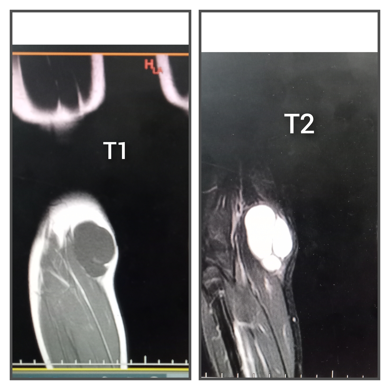
MR
T1 low signal
T2 high signal
Grey scale
- well-defined cyst with a 'neck' at its deepest extent
- extending into the joint space between the semimembranosus tendon and the medial head of the gastrocnemius
- identification of a fluid-filled structure at the posteromedial knee is suggestive of a popliteal cyst, but identification of the 'neck' between the tendons is necessary for a definitive diagnosis
- anechoic, but may contain internal debris
Leave a Reply