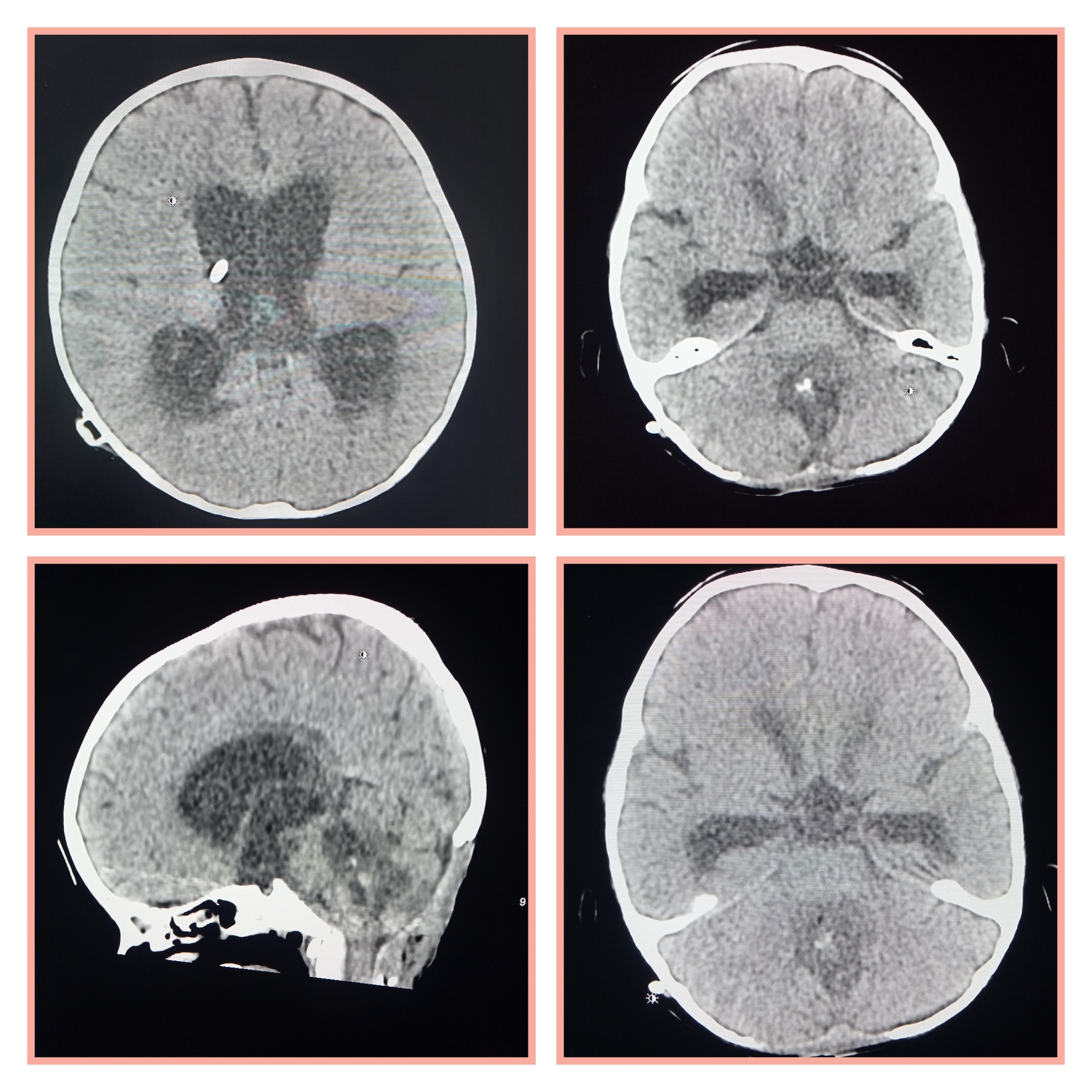
Imaging features of medulloblastoma
CT features of medulloblastoma
- medulloblastomas often appear as a mass arising from the vermis
- effacement of the fourth ventricle / basal cisterns and obstructive hydrocephalus.
- hyperdense on CT
- cysts formation/necrosis is common
- Enhancement
MRI
T1
hypointense to grey matter
T1 C+ (Gd)
enhancely heterogenously
T2/FLAIR
hyperintense to grey matter
heterogeneous due to calcification, necrosis and cyst formation
surrounding oedema is common
DWI/ADC
high DWI signal ("restricted diffusion") - due to their hypercellularity
MR spectroscopy
elevated choline
decreased NAA
Leave a Reply