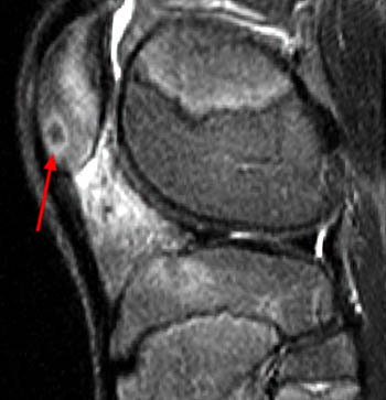
Magnetic resonance imaging
• T1: Nidus is isointense to muscle.
• T2: Nidus shows intermediate to high signal.
Peak contrast enhancement occurs during the arterial phase
with early partial washout. Extensive bone marrow
edema may be present, which can obscure the nidus.
• Bone scan: Uptake of radioactive tracer is increased. May
appear as a small focus of intense activity surrounded by
a larger area of increased activity.
• Angiography: Vascularity is increased in the region of the
lesion with dense enhancement of the nidus. A feeding
vessel may be identified.
osteoid osteoma
cc radsource, paeds thieme
Leave a Reply