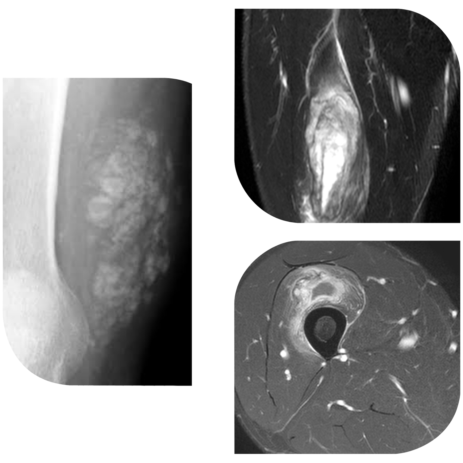
ovoid region of soft-tissue swelling slightly hyperintense to muscle (arrows) is seen on the T1-weighted coronal image. Subtle areas of peripheral low signal intensity (arrowheads) are seen within the lesion. (2B,C) The fat-suppressed proton density-weighted axial and T2-weighted coronal images reveal a hyperintense lesion within the vastus intermedius muscle (arrows) with a small region of central fluid signal intensity (asterisks). The abnormality demonstrates lace-like intramuscular enhancement (arrows) with thicker enhancement surrounding the central fluid (asterisk) on the (2D) fat-suppressed T1-weighted post-contrast view. Peripheral calcifications (arrows) are readily apparent on the corresponding (2E) lateral radiograph of the thigh.
Case courtesy: https://radsource.us/myositis-ossificans/
Leave a Reply